What is a digital dental X-ray?
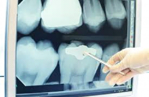
In a digital dental X-ray, a sensor is placed inside your mouth, which then projects an image onto the attached computer screen. Unlike traditional X-ray which takes time to build and is printed out on film, digital dental X-ray exists digitally and is formed in less than a few minutes.
The process of digital dental x-ray is quite simple — as soon as the sensor is placed at the right position inside the mouth, the information is sent directly to the attached computer. Either we can instantly view the inside of the person’s mouth on the screen, or we can capture images using a specialized scanner. It is known that digital dental x-rays are more efficient and safer than traditional film x-rays.

Why do you need a digital X-ray?
When the dentist examines your mouth in the clinic, he or she can only have a partial view of it. There are many parts of the inside of your mouth that are not visible to them. For an in-depth oral examination, the dentist needs to view every little corner of your mouth. Dental X-rays are needed to properly monitor the condition of your teeth, roots, and jaw placement, and assist in finding and treating dental problems early in their development. These problems are usually not detected in an oral exam and require X-rays to be checked.
There are many problems a digital dental X-ray can detect, such as tooth decay, changes in the bone or root canal due to infection, condition and position of teeth for implants, braces, dentures, or other dental procedures, position and angle of wisdom teeth, internal tooth fractures, infections, cysts and some types of tumors.

What are the advantages of digital X-rays?
- Digital dental X-rays produce very less radiation compared to traditional X-rays, which makes them safer to use.
- Digital dental X-rays are formed instantly on the screen, which helps save time for the dentist and the patient.
- Once the image in the dental x-ray is formed on the screen, it can be adjusted, zoomed in, zoomed out, enlarged, etc., which makes it easier to spot and identify problems such as small cavities, a fracture in the tooth, etc.
- Digital dental X-rays can be digitally stored, so they are easy to access for both the doctor and the patient. They don’t have to be carried everywhere.
- With the help of digital dental X-rays, dental problems can be easily explained to the patient right then and there.
- Digital dental X-rays are environmentally and cost-friendly.
How often do you need digital X-rays?
The frequency of the need for digital dental X-rays depends upon the patient and the kind of treatment they are undergoing. Usually, healthy patients don’t need to get digital X-rays that often. But if the patient goes through dental problems quite often, digital dental X-rays may be suggested often as there is more risk of developing new problems. Children also need to get dental X-rays more often than adults. This is because they are more likely to go through dental problems due to their growing age. Many children develop cavities growing up, have problems with wisdom teeth, etc.
Sometimes, special circumstances also require digital dental X-rays. For example: if a person has good dental health but has recently had an injury in their jaw or if they experience a sudden toothache.
Types of digital X-rays:
There are mainly two types of digital dental X-rays, that have some subtypes of their own:
1. Intraoral X-rays:
These are the kind of digital dental X-rays that are taken inside your mouth. There are a few types of intraoral X-rays:
Bite-Wing X-rays:

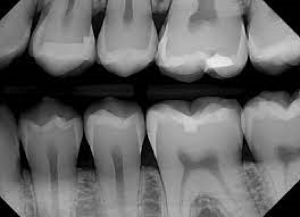
The most common kind of X-ray, in which you have to bite down on a sensor, Bite-Wing X-rays produce images that show the upper and lower teeth in one area of the mouth. Each bite wing shows a tooth from its crown to the level of the supporting bone. This way, the dentist is able to identify any decay in between teeth, check the condition of bone around teeth, and determine the proper fit of a crown.
Periapical X-rays:

This kind of x-ray shows the whole tooth, from its crown to the tip of the tooth root, and the bone surrounding the tooth. Each periapical X-ray displays the full tooth dimension and includes all the teeth in one portion of either the upper or lower jaw. This way, the dentist is able to detect any abnormalities in the root structure and the surrounding bone structure.
2. Extraoral X-rays:
These are the kind of digital dental X-rays that are taken outside your mouth. One type of extraoral X-ray is:
Panoramic X-rays:

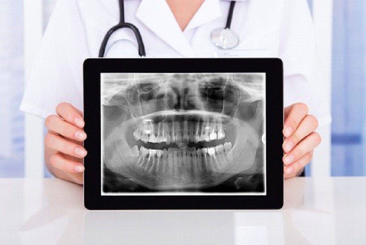
This kind of x-ray is taken by a machine that rotates around your head and provides a detailed image of all your teeth in your upper and lower arch. Looking at that image, the dentist can see fully emerged and emerging teeth. They can also easily identify impacted teeth, and jaw problems, and diagnose tumors.
At Dental RI, you will find that all our digital x-ray equipment is exceptionally well maintained. We make sure to provide the suitable service for the problems our patient is undergoing, and always take every precaution to keep them safe during their treatments.
